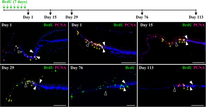
Figure 5.

Identification and localization of putative slow‐cycling stem cells in the peripheral retina of P. vitticeps. The top schematic drawing depicts the experimental strategy and BrdU pulse‐chase time points (black arrows). Retinal tissues were collected in adult (>2‐years old, top left panel) and/or juvenile (<1‐year old, other panels) P. vitticeps at 1, 15, 29, 76, or 113 days after the first week of BrdU feeding, and sections from the peripheral dorsal retina were processed for double immunohistochemistry against BrdU (green) and PCNA (red). Solid arrowheads delimitate the retinal peripheral margin, and open arrowheads indicate the boundary between the monolayered ciliary epithelium and the RCJ. The localization and number of yellow asterisks reflect the position and relative abundance of BrdU/PCNA double‐positive cells, respectively, in the ciliary epithelium, RCJ, and retinal margin. Only BrdU labeling is shown after 76 days of chase to better highlight the diluted BrdU‐labeled cells scattered outside the RCJ within both the ciliary body and the retina (green arrows). Cell nuclei are counterstained with DAPI (blue). Scale bars, 100 μm
