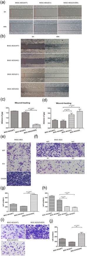
Figure 3.

CircFNDC3B promoted migration and invasion. (a,b) A wound‐healing assay was performed to evaluate the migration ability of MGC‐803 and BGC‐823. MGC‐803 cells were transfected with circFNDC3B vector (OVER) or empty vector (EV) for 48 hr. BGC‐823 cells were transfected with siRNA (S1, S2) or negative control siRNA (NC) for 48 hr. A line was drawn from one end of the well to the other. The wound‐healing process was recorded by microscopy at 100× magnification. The red line indicates the measured distance after cell migration (WT: wild‐type; EV: empty vector; OVER: overexpressing circFNDC3B; NC: negative control; siRNA: S1, S2). (c,d) The migration ability was evaluated by the red line distance of the wound‐healing gap. Quantitative results showed that the wound gap was obviously shortened in MGC‐803 cells transfected with the circFNDC3B vector and that silencing circFNDC3B suppressed cell migration in BGC‐823 cells (WT: wild‐type; EV: empty vector; OVER: overexpressing circFNDC3B; NC: negative control; siRNA: S1, S2). (e,f) Transwell assays were performed to evaluate the invasion ability of MGC‐803 and BGC‐823 cells after overexpression and silencing of circFNDC3B. MGC‐803 cells were transfected with circFNDC3B vector (OVER) or empty vector (EV) for 48 hr. BGC‐823 cells were transfected with siRNA (S1, S2) or negative control siRNA (NC) for 48 hr. The cells were trypsinized, resuspended, and seeded on the upper chamber of well‐coated ECM gel. After 24 hr, the invaded cells on the bottom of the well were fixed and stained. The invaded cells stained by crystal violet were calculated as the degree of invasion. Three different areas of each well were randomly selected and observed under a microscope at 200× magnification. The results showed that MGC‐803 cells transfected with the circFNDC3B vector invaded more than the empty vector cells, whereas silencing circFNDC3B in BGC‐823 cells caused them to invade less than BGC‐823 cells with negative control (WT: wild‐type; EV: empty vector; OVER: overexpressing circFNDC3B; NC: negative control; siRNA: S1, S2). (g,h) Quantitative results showed that overexpressing circFNDC3B promoted cell invasion in MGC‐803 cells and that silencing circFNDC3B suppressed cell invasion in BGC‐823 cells (WT: wild‐type; EV: empty vector; OVER: overexpressing circFNDC3B; NC: negative control; siRNA: S1, S2). (i,j) BGC‐823 cells were transfected with the circFNDC3B vector, and results showed that circFNDC3B indeed promoted cell migration and invasion (WT: wild‐type; EV: empty vector; OVER: overexpressing circFNDC3B). Finally, we validated that circFNDC3B could promote cell migration and invasion. Data were expressed as the mean ± SEM and were analyzed by independent samples t test, n = 3. *p < 0.05, ** p < 0.01, *** p < 0.0001. ECM: extracellular matrix; FNDC3B: fibronectin type III domain‐containing protein 3B; SEM: standard error of the mean; siRNA: small interfering RNA [Color figure can be viewed at wileyonlinelibrary.com]
