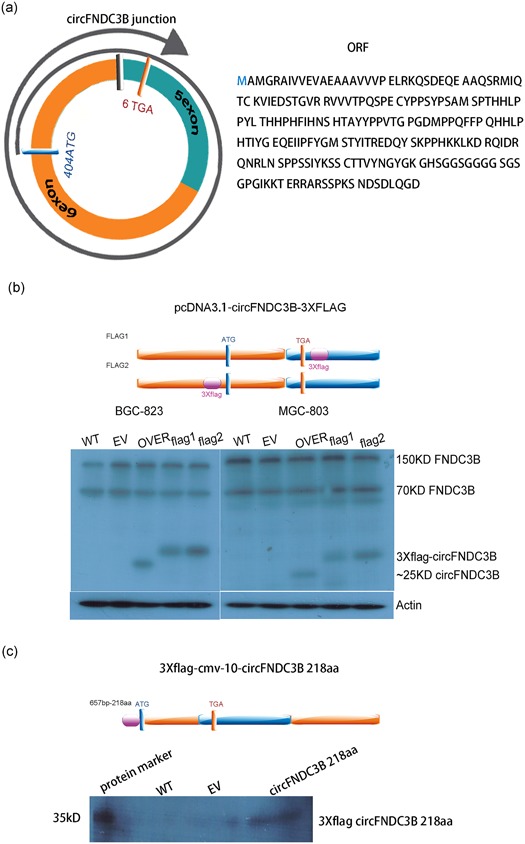
Figure 7.

CircFNDC3B has translational protein activity. (a) The figure shows start codon and stop codon positions, 2r+8 rolling circle translation format, and protein sequence information (blue ring: 5 exon; orange ring: 6 exon; black square: junction; blue square: start codon; orange square: stop codon). (b) The circFNDC3B translational protein was detected by inserting a 3× flag–tag at both ends of the initiation codon and the stop codon of the circFNDC3B cyclization vector pcDNA3.1 circFNDC3B mini vector. MGC‐803 cells were transfected with circFNDC3B vector (OVER) or empty vector (EV) or vector with flag–tag for 48 hr. BGC‐823 cells were transfected with circFNDC3B vector (OVER) or empty vector (EV) or vector with flag–tag for 48 hr. Western blot results showed that circFNDC3B could translate approximately 25 kD peptide against the FNDC3B‐specific antibody and could also detect the 150 and 70 kD FNDC3B variants (purple square: 3× flag–tag; blue ring: 5 exon; orange ring: 6 exon; black square: junction; blue square: start codon; orange square: stop codon). (c) A linear expression vector was synthesized and constructed according to the circFNDC3B rolling circle translation sequence. MGC‐803 cells were transfected with p3× flag‐CMV‐10‐circFNDC3B or p3× flag‐CMV‐10 empty vector (EV) for 48 hr. Western blot results showed that circFNDC3B has translational protein activity against flag–tag antibodies (purple square: 3× flag; blue ring: 5 exon; orange ring: 6 exon; black square: junction; blue square: start codon; orange square: stop codon). FNDC3B: fibronectin type III domain‐containing protein 3B [Color figure can be viewed at wileyonlinelibrary.com]
