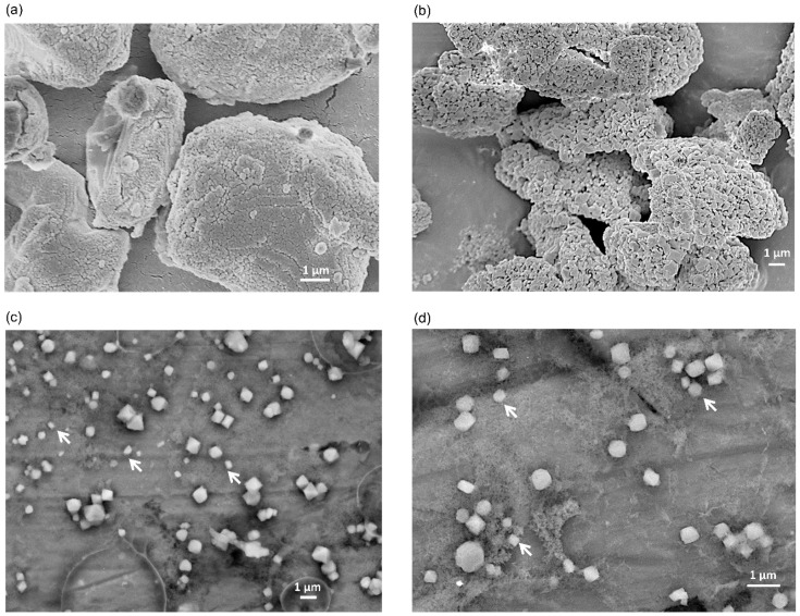
Figure 3.
SEM images of BiSCaO-6 dry powder and dispersion. The surface structure of each dry powder of BiSCaO-6 immediately after opening at 10,000-fold magnification (a) and BiSCaO-6 placed under high humidity at 37 °C for seven days at 5000-fold magnification (b) were observed with SEM images of a field-resolved scanning electron microscope. Cryo-SEM observations were performed on BiSCaO-6 dispersions (0.2 wt% BiSCaO-6, 0.12 wt% Na2HPO4) two days after adjustment at 5000-fold magnification (c) and 10,000-fold magnification (d). Allows indicate nano particles.

