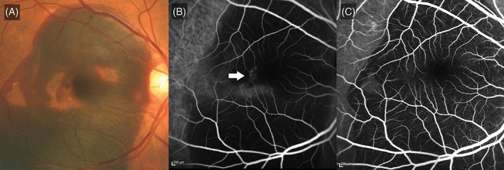
Figure 3.

Massive submacular haemorrhage with blocked fluorescence. A, Colour fundus photograph illustrating massive subretinal haemorrhage. Pigment epithelial detachments are seen within the area of haemorrhage. B, Indocyanine green angiogram illustrating hyperfluorescence (white arrow) which is partially masked by the thick layer of haemorrhage. C, Fluorescein angiogram showing blocked fluorescence as a result of the haemorrhage
