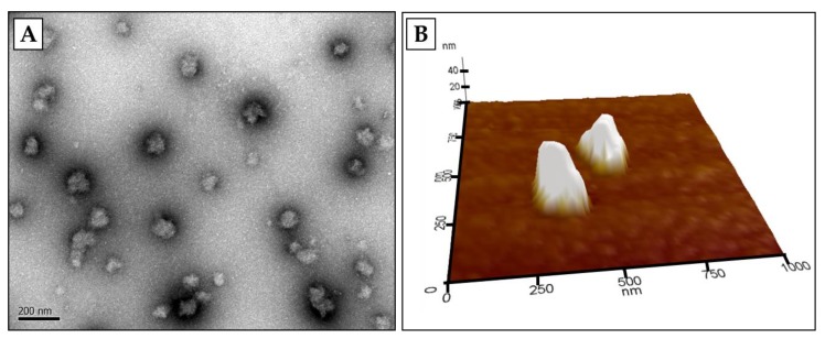
Figure 2.
Microscopic images of β-lg nanoparticles observed in transmission electron microscopy (TEM) (A) and atomic force microscopy (AFM) (B). β-lg nanoparticles were prepared using the modified ionic gelation method described in Ha et al. [24]. This figure is original and has not been previously published. Scale bar = 200 nm.

