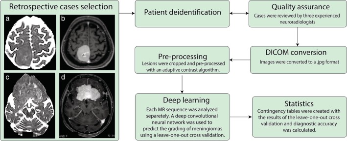
Figure 1.

Schematic representation of the workflow used in this retrospective study. Axial (a) ADC Maps, (b) PCT1W images obtained in a 58‐year‐old woman showing a WHO Grade I right fronto‐parietal meningioma. Axial (c) ADC Maps, (d) PCT1W images obtained in a 72‐year‐old woman showing a WHO Grade II bilateral fronto‐basal meningioma.
