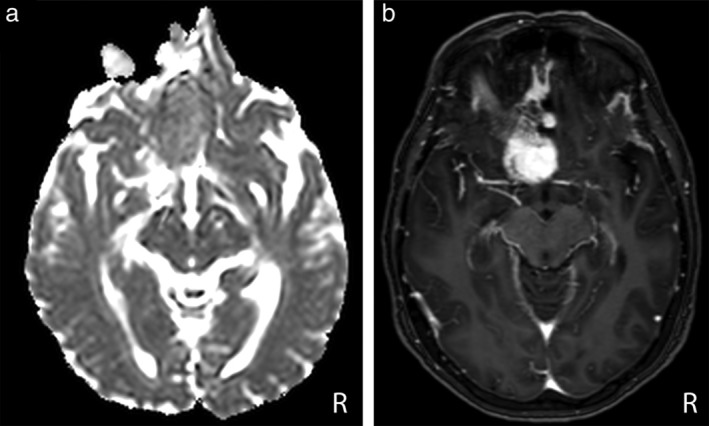
Figure 5.

Axial ADC map (a) and PCT1W images (b) in a 59‐year‐old woman with a Grade I anterior clinoid meningioma. The lesion was misclassified as a WHO Grade II–III lesion by the IncV3‐ADC model, whereas it was correctly classified as a WHO Grade I lesion by the IncV3‐PCT1W model.
