Figure 7.

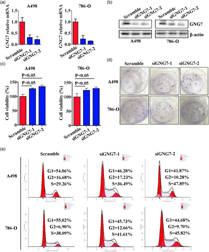
Loss of GNG7 facilitated cell proliferation by increasing G2/M cell‐cycle phase. A498 and 786‐O cells were transfected with oligo siGNG7 or negative control oligo small interfering RNA (siRNA). After 48 hr, cells lysates were collected and expression of GNG7 was analyzed by real‐time quantitative polymerase chain reaction (a) and western blot analysis (b). (c) MTT assay was used to detect cell viability in A498 and 786‐O cells transfected with GNG7 siRNA. (d) The colony formation ability in A498 and 786‐O cells was visualized by crystal violet staining. (e) Flow cytometric analysis was used to detect cell cycle of A498 and 786‐O cells after transfection. p < 0.05 was considered statistically significant [Color figure can be viewed at wileyonlinelibrary.com]
