Figure 1.
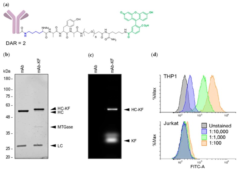
MTGase site specifically labels engineered anti-CD33 mAb. (a) Structure of mAb-KF peptide conjugate. Conjugated lysine is highlighted in blue and fluorescein is highlighted in green; (b) UV protein stained SDS-PAGE gel of unconjugated mAb and mAb-KF. Molecular weight markers are in kilodaltons (kDa). HC—heavy chain, LC—light chain, HC-KF—heavy-chain-KF conjugate, MTGase—microbial transglutaminase; (c) FITC epifluorescence image of the same SDS-PAGE gel. KF—free KF peptide; (d) Flow cytometry of mAb-KF stained THP1 (CD33-positive) and Jurkat (CD33-negative) cells. Following spin column cleanup, mAb-KF conjugate was directly diluted in stain buffer at the indicated ratios: blue—1:10,000, green—1:1,000, orange—1:100. Unstained cells are represented in gray. All plots are represented as percent maximum cell count.

