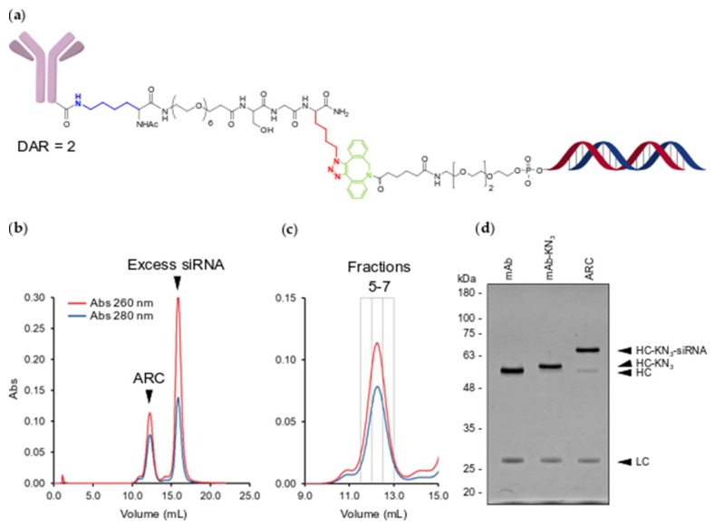
Figure 3.
Conjugation and purification of ARC. (a) Structure of mAb-KN3-DBCO-TEG-siRNA conjugate. Conjugated lysine is highlighted in blue. Conjugated triazole (azidolysine origin) is highlighted in red. Conjugated DBCO moiety is highlighted in green; (b) SEC analysis of the crude ARC conjugation reaction. UV absorbance at 260 nm is traced in red and absorbance at 280 nm is traced in blue; (c) SEC purification of ARC. Gray boxes indicate the elution volumes of collected fractions (5–7); (d) UV protein stained SDS-PAGE gel of unconjugated mAb, mAb-KN3 and ARC. Molecular weight markers are in kilodaltons. HC—heavy chain, LC—light chain, HC-KN3—heavy-chain-KN3 conjugate, HC-KN3-siRNA–heavy-chain-KN3-siRNA conjugate.

