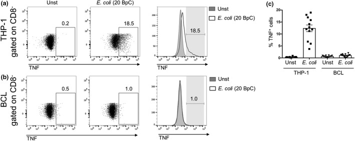
Figure 2.

THP‐1 cells but not B cells produce TNF upon bacterial stimulation. THP‐1 cells (a) or BCLs (b) were co‐cultured with enriched CD8 T cells for 20 h in the absence (Unst) or presence of 20 BpC E. coli. THP‐1 cells or BCLs defined by gating on CD8 negative cells were analyzed for TNF expression after intracellular cytokine staining. Numbers next to gates represent percentage of positive cells. (c) Mean ± s.e.m. TNF + THP‐1 or BCLs are shown from 12 donors pooled from three independent experiments.
