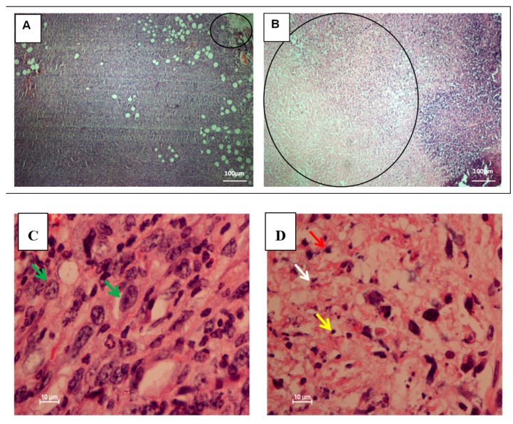
Figure 2.
Hematoxylin and eosin (H&E) staining of breast tumors in xenografts of mice induced by 4T1 cells. The untreated 4T1-injected mice breast tumors show focal areas of necrosis/late apoptosis making up about 5%–10% of the lesions (Panel A). Treated tumors demonstrated large areas of necrosis/late apoptosis at the central part of the tumors about 70% and about 60% of the lesions, respectively (Panel B). Higher magnification of untreated 4T1-injected mice breast tumors show mostly viable tumor cells indicated with pleomorfic vesicular nuclei and hyperchrome prominent nucleoli (green arrow) (Panel C). Higher magnification of citral-treated 4T1 breast tumors show large areas of necrosis/late apoptosis indicated with increased number of apoptotic cells (red arrow), cellular debris (white arrow), and nuclear dust (yellow arrow) (Panel D), (20×, 100×, scale bar represents 100 and 10 µm in length). The data were generated from six fields per slide, with four slides analyzed from each tumor, and 3 tumors examined from each group. (n = 6).

