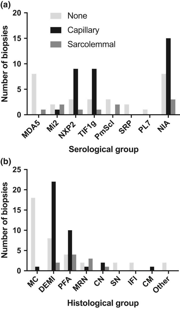
Figure 5.

Correlation of complement attack complex (C5b‐9) deposition pattern with serological and histological groups across the study cohort. The results of immunohistochemical staining for C5b‐9 were available for 76 patients after restriction to patients not on steroid treatment at the time of biopsy. Patient numbers whose biopsies showed capillary complement deposition, sarcolemmal complement deposition, or neither are plotted against (a) serological and (b) histological groups. For statistical analysis cases with sarcolemmal membrane attack complex deposition and without any deposition were merged into a single category in order to ensure meaningful group sizes. For results see text. MC, minimal change; DEMI, diffuse endomysial macrophage infiltrates; PFA, perifascicular atrophy; MRN, macrophage rich necrosis; CN, clustered necrosis; SN, scattered necrosis; FI, fibre invasion; CM, chronic myopathic change. NIA, no identifiable autoantibodies.
