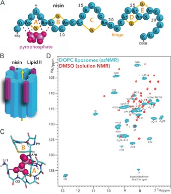
Figure 5.

A) Nisin features five thioether rings, of which rings A and B are critical for binding lipid II. B) Nisin and lipid II form a defined pore that spans the bacterial plasma membrane. C) The pyrophosphate cage structure as determined in DMSO.7 The thioether rings A and B of nisin are annotated. D) Overlay of 2D NH spectra of the nisin⋅lipid II complex in DOPC (blue) and in DMSO (red). The ssNMR spectrum was measured at 950 MHz (1H frequency) and 60 kHz magic‐angle spinning. The solution NMR spectrum in DMSO was published previously.7 (A) and (D) reproduced from ref. 30. Copyright: 2018, the authors.
