Figure 1.

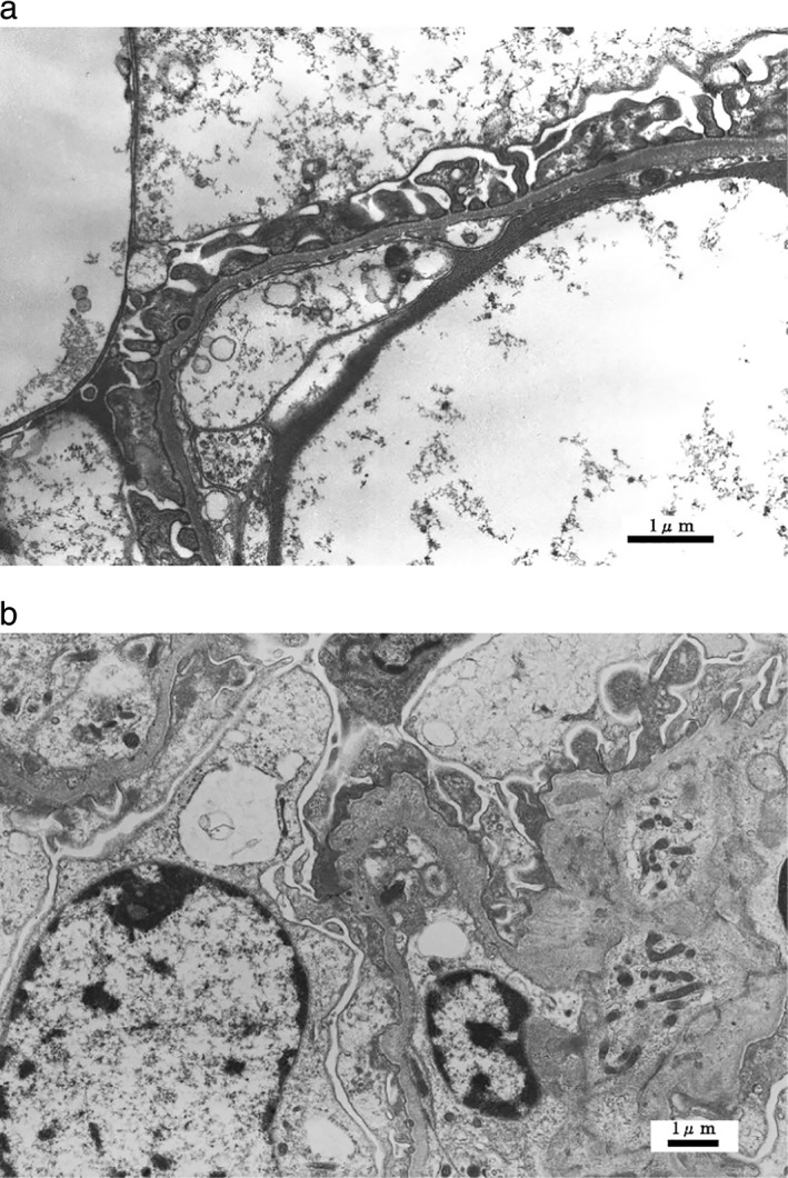
Typical glomerular basement membrane (GBM) changes on electron microscopy. (a) Thinning of the GBM (×6000), (b) Lamellation of the GBM (×6000).

Typical glomerular basement membrane (GBM) changes on electron microscopy. (a) Thinning of the GBM (×6000), (b) Lamellation of the GBM (×6000).