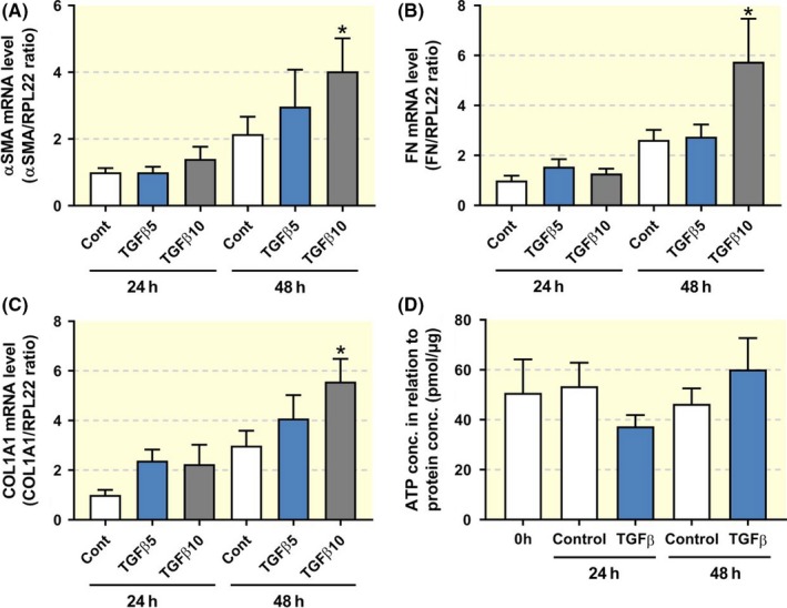
Figure 6.

Expression of fibrosis markers in human PCKS. PCKS were exposed to TGF‐β (5 or 10 ng/ml) for 24‐48 h. (A‐C) Gene expression was studied by qPCR. Relative expression was calculated using the reference gene RPL22 (n = 4‐5). (D) Viability of PCKS after treatment with 10 ng/ml TGF‐β, assessed by ATP content of the slices (n = 5‐7). Data are presented as mean ± SEM. *P < 0.05
