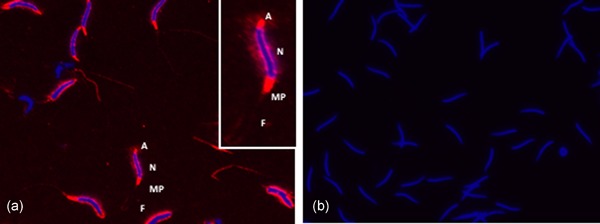
Figure 3.

Immunofluorescence of SPINK2 in chicken sperm. (a) Expression of SPINK2 on the sperm. (b) No signal was detected in the control slide (incubated with the secondary antibody only). Red color: SPINK2 localization in acrosome (a), midpiece region (MP) and flagella (F); blue color: staining of the nucleus (N)
