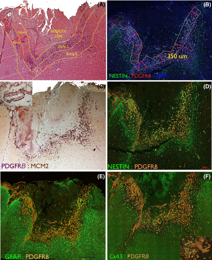
Figure 1.

Intracranial recording (ICR) injury at low power. (A) A subacute lesion (13 days post injury) involving the cortical surface which represents one of the larger foci of injury in the series. H&E section highlighting the main regions used for qualitative evaluation and delineation of the Zones for study. The Necrotic core (Zone 0) contains mainly nonviable material and macrophages. Zone 1 is the rim of viable material surrounding the core and contains reactive cells and vessels and Zone 2 surrounds Zone 1. (B) The same case showing the superimposed measured Zone 1 with a width of 350 microns which was tiled for image analysis. (C) PDGFRβ chromogenic stained section (purple) with MCM2 labelling of nuclei (brown, visible in inset) in the same case to highlight the labelling in Zone 1 of vessels and in (D). With immunofluorescent labelling for PDGFRβ with nestin highlighting capillaries and reactive glial cells in Zone 1. (E) GFAP with PDGFRβ shows the relative compartmentalization of labelling at low power with the majority of GFAP positivity external to the PDGFRβ. (F) Cx43 with PDGFR β at low power and shown in the inset at higher magnification, with a close relationship between PDGFRB pericytes surrounding Cx43‐positive endothelium. Higher magnifications for C are shown in Figure 4, D in Figure 2, E in Figure 3 and F in Figure 5. Bar is 700 microns.
