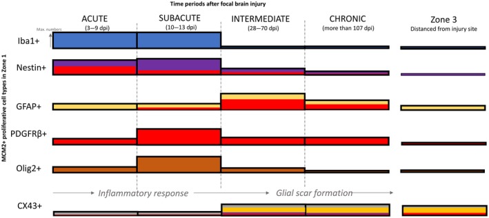
Figure 6.

Summary schematic illustrating the expression of different proliferative cell types around the lesion at various intervals post injury. This is based on the data in current and previous study 5. The height of the bar refers to the level of expression or numbers of dividing cell types. Immediately after injury, the number of dividing Iba1+ microglia and nestin+ expressing cells are upregulated around the lesion, reaching maximal numbers around 2 weeks post lesion. At 2 weeks, an increased number of dividing PDGFRβ+ and Olig2+ expressing cells were also observed around the lesion. After a month post lesion, the number of Iba+ microglia and Olig2+ oligodendrocytes decreased dramatically, reaching similar number of cells found in Zone 3 (normal level). In contrast, many dividing GFAP + astroglia were observed only after 1 month after injury. A higher number of MCM2+/GFAP + cells were still found around lesion after 4 months post lesion, together with few nestin+ and PDGFRβ+ cells. The level of co‐expression between PDGFRβ+ and nestin (red bars within nestin+) was maximally observed a week after post lesion and gradually decrease with time. In contrast, the PDGFRβ+ and GFAP was maximally observed 2 months after lesion. The expression of CX43 was significantly reduced from normal level, immediately following injury, but increased after a month, coinciding with the upregulation of GFAP. Some PDGFRβ+and nestin+ expressing cells in Zone 1 were found to express CX43 1 month after lesion (Bar colours ‐ Blue, Iba1; purple, nestin; yellow, GFAP; red, PDGFRbeta; brown, olig2 and grey, Cx43).
