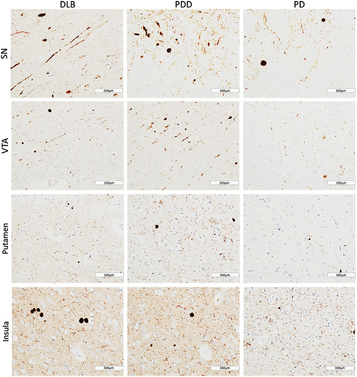
Figure 1.

α‐synuclein pathology distribution . Photomicrographs of α‐synuclein pathology distribution in substantia nigra (SN), ventral tegmental area (VTA), putamen and insula in DLB (case #4, LB Braak stage 6), PDD (case #26, LB Braak stage 6) and PD (case #34, LB Braak stage 4). α‐synuclein immunohistochemistry with KM51 antibody, ×20. Scale bar 300 µm.
