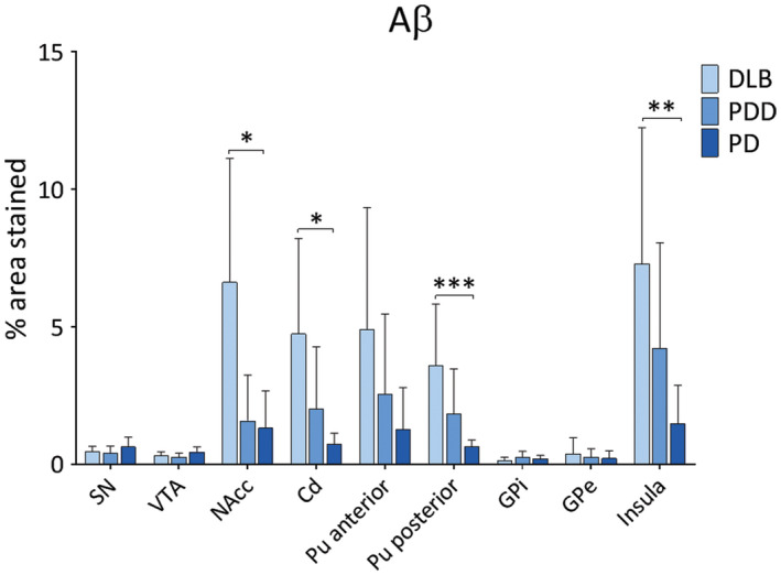
Figure 6.

Amyloid‐beta pathology in LBD . Aβ pathology (% area stained) was assessed in substantia nigra (SN), ventral tegmental area (VTA), nucleus accumbens (NAcc), caudate (Cd), anterior and posterior putamen (Pu), globus pallidus internus (GPi) and externus (GPe) and insula between DLB, PDD and PD groups. *P < 0.05, DLB vs. PD; **P < 0.01, DLB vs. PD; ***P < 0.001, DLB vs. PD.
