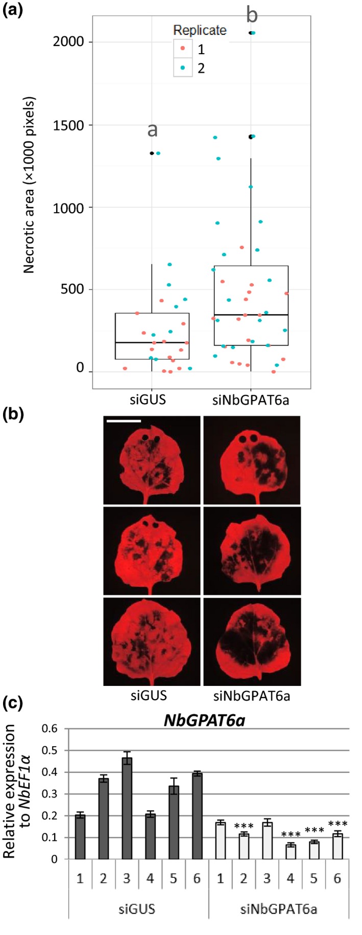
Figure 3.

siNbGPAT6a‐mediated virus‐induced gene silencing (VIGS) results in stronger Nicotiana benthamiana leaf necrosis upon Phytophthora infestans infection. (a) Leaf necrotic area 5 d postinoculation (dpi) with P. infestans, as quantified by lack of red Chl fluorescence, two replicates combined (P = 0.0127). Data points for each replicate are denoted by different colours. Horizontal lines represent median and upper and lower quartiles. Whiskers extend to data points that are < 1.5 × interquartile range away from upper/lower quartile. Lower case letters ‘a’ and ‘b’ indicate significant differences between the means as compared using Student's t‐test (P < 0.05). (b) Representative UV images of leaf necrosis quantified in (a). Scale bar, 30 mm. Holes at the leaf tip in some images are the result of tissue samples taken for quantification of transcript abundance (these areas were excluded from necrotic area quantification). (c) Relative expression of NbGPAT6a (to NbEF1α) following VIGS using siNbGPAT6a or siGUS (control). Error bars represent ± SE of the mean of three biological replicates. Student's t‐test was used to compare mean relative expression in siNbGPAT6a samples with the lowest of control samples. *, P < 0.05; **, P < 0.01; ***, P < 0.001.
