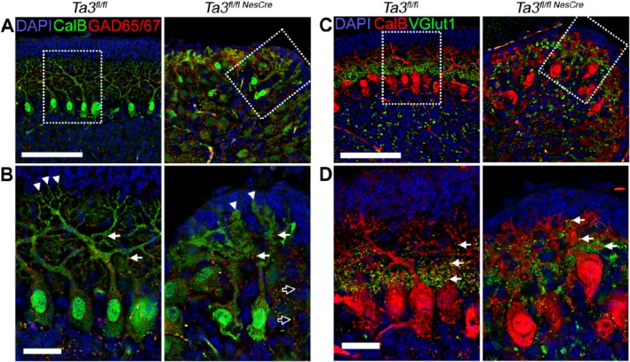
Figure 5.

Inhibitory and excitatory synapses in Purkinje neurons on Talpid3 mutant and wild‐type mice. (A, B) P10 wild‐type and Talpid3 mutant cerebellar sections immunostained with anti‐GAD‐65/67 for inhibitory synapses. The box indicates region of greater magnification (B). (C, D) P10 wild‐type and Talpid3 mutant cerebella immunostained with anti‐VGlut1 for excitatory synapses at Purkinje cell dendrites. The box indicates region of greater magnification (D). CalB, calbindin; GAD, glutamic acid decarboxylase; VGlut1, vesicular glutamate transporter 1. Scale bar: 100 μm (A, C); 25 μm (B, D). White arrow heads‐dendritic spines; white arrows ‐ synapses; Outlined black arrows ‐ abnormally placed synapses.
