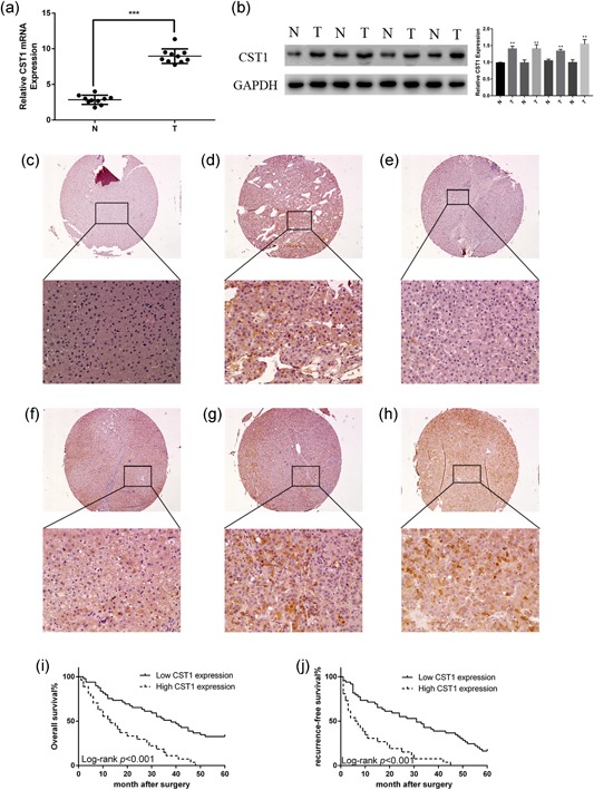
Figure 1.

CST1 was overexpressed in human HCC and predicted poor prognosis. (a) mRNA levels of CST1 in different HCC tissues and matched adjacent normal liver tissues. (b) Protein levels of CST1 in different HCC tissues and matched adjacent normal liver tissues (left). Gray value ratio (right). (c) Negative control in normal liver tissue. (d) Positive cytoplasmic expression in liver tissue. (e) Negative control in HCC. (f) Weak positive cytoplasmic expression in HCC. (g) Moderate positive cytoplasmic expression in HCC. (h) Strong positive cytoplasmic expression in HCC. (i) Kaplan–Meier analysis of overall survival in patients with HCC. (j) Kaplan–Meier analysis of recurrence‐free survival in patients with HCC. Scale bar = 100 μm (top) and 500 μm (bottom). Data are presented as means ± standard deviations of three independent experiments. GAPDH: glyceraldehyde 3‐phosphate dehydrogenase; HCC: hepatocellular carcinoma; mRNA: messenger RNA. *p < .05; **p < .01; and ***p < .001 [Color figure can be viewed at wileyonlinelibrary.com]
