Figure 4.

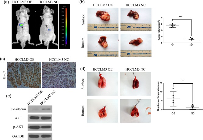
CST1 promoted HCC growth and metastasis in vivo. (a) Images of the orthotopic model were acquired using bioluminescence imaging. (b) CST1 overexpression increased HCCLM3 cell growth in the orthotopic model in nude mice. Tumor volumes were calculated. (c) Images of immunohistochemical detection of Ki‐67 in orthotopic HCC tissues. Scale bar = 500 μm. (d) CST1 overexpression promoted HCCLM3 cell metastasis to the lung. The number of nodules transferred was counted. (e) The levels of E‐cadherin, phospho‐AKT, and total AKT in the lung metastasis model were measured by western blot analysis. Data are presented as means ± standard deviations of three independent experiments. GAPDH, glyceraldehyde 3‐phosphate dehydrogenase; HCC, hepatocellular carcinoma; NC, negative control. *p < .05; **p < .01; and ***p < .001 [Color figure can be viewed at wileyonlinelibrary.com]
