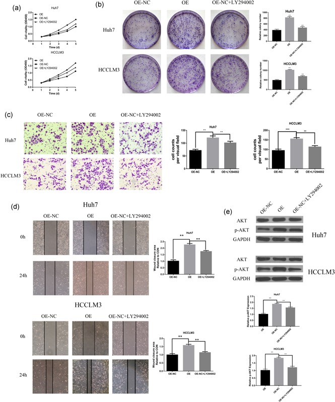
Figure 5.

PI3K/AKT signaling pathway involved in CST1‐induced HCC progression. (a) LY294002 decreased the CST1‐induced proliferation curves. (b) Colony formation assays (left) demonstrated that LY294002 could reverse cell proliferation induced by CST1, Number of colonies counted (right). (c) Representative images from the transwell invasive assays, LY294002 decreased the CST1‐induced invasion of Huh7 and HCCLM3 cells. The number of invaded cells was counted. (d) Representative images from the wound‐healing assays, LY294002 decreased the CST1‐induced migration of Huh7 and HCCLM3 cells. The wound‐closure area was calculated. (e) Western blot analysis (left) of p‐AKT expression after LY294002 treatment with Huh7 and HCCLM3 cells. Gray value ratio (right). GAPDH: glyceraldehyde 3‐phosphate dehydrogenase; HCC: hepatocellular carcinoma; NC: negative control. *p < .05; **p < .01; and ***p < .001 [Color figure can be viewed at wileyonlinelibrary.com]
