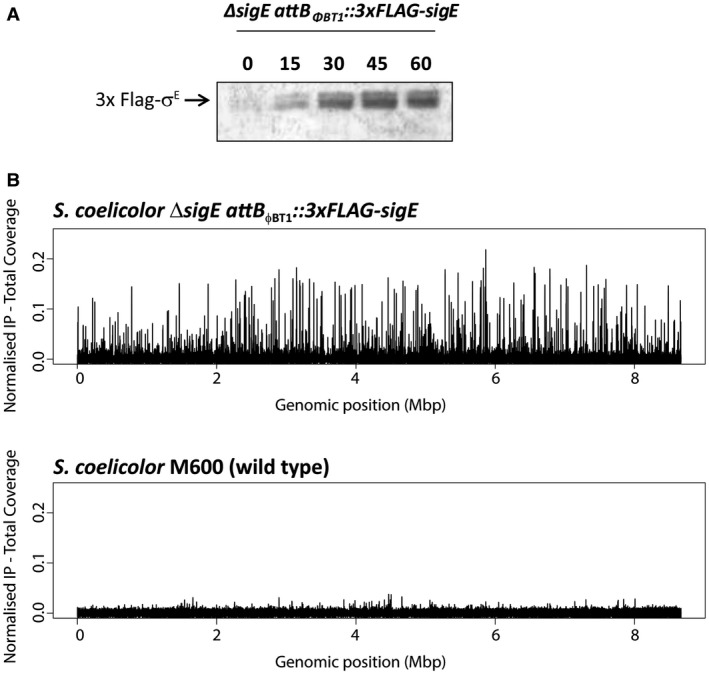
Figure 2.

A. Western blot analysis of S. coelicolor ΔsigE attBΦBT1::3 × FLAG‐sigE grown in NMMP liquid cultures and sampled after 0, 15, 30, 45 and 60 minutes treatment with 10 µg/ml vancomycin. Total protein (10 µg) was loaded per lane and 3 × FLAG‐σE was detected using anti‐σE polyclonal antibody. B. Chromosome‐wide distribution of σE‐binding sites in S. coelicolor identified by ChIP‐seq analysis. ChIP‐seq was conducted using M2 anti‐FLAG antibody on the ΔsigE attBΦBT1::3 × FLAG‐sigE strain after 30 minutes treatment with 10 µg/ml vancomycin. The wild‐type strain (expressing non‐tagged σE from the native locus) analysed under the same conditions was used as a negative control.
