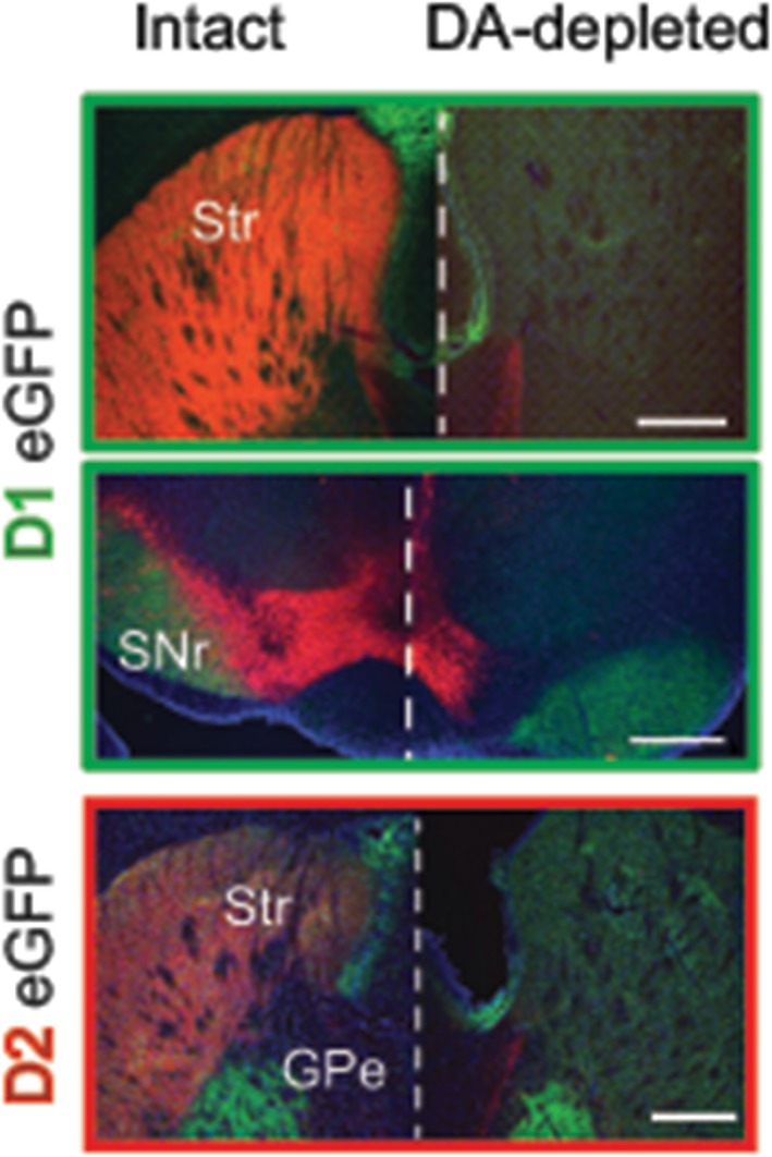
Figure 2.

Immunohistochemistry of control and dopamine‐depleted brains. Coronal brain slices of intact and DA‐depleted D1eGFP+ and D2eGFP+ mice illustrate DA neurons and axons by tyrosine hydroxylase (TH) staining (red); positive expression of fluorescent protein (green) and the presence of cell bodies stained with 4,6‐diamidino‐2phenylindole, dihydrochloride (DAPI) (blue). GPe: external globus pallidus; Scale bars: 1 mm; Str: striatum; SNr: substantia nigra pars reticulata
