Figure 1.

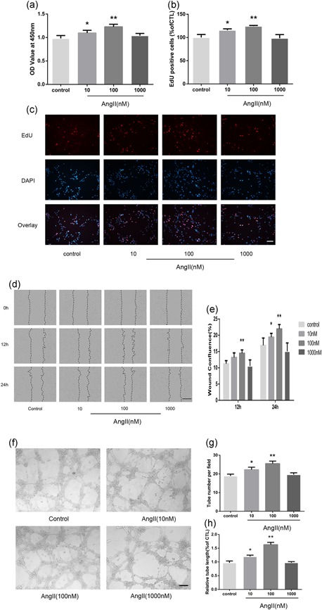
AngII induces human microvascular endothelial cell (HMEC) proliferation, migration, and capillary tube formation. (a) A Cell Counting Kit‐8 assay was performed to evaluate cell proliferation after 24 hr in corresponding serum‐free medium (n = 6). (b, c) EdU‐labeling analysis showed the fluorescence images of HMECs stimulated with 0, 10, 100, or 1,000 nM AngII for 24 hr (EdU, red fluorescent signals; DAPI, blue signals; ×100). (c) The percentage of DAPI‐positive cells indicated the quantification of EdU‐positive HMECs (n = 3; b). (d) HMECs monolayers were scratched and incubated with 0, 10, 100, or 1,000 nM AngII. Microscopic images were taken at 0, 12, and 24 hr using the IncuCyte ZOOM Live‐Cell Analysis System (Essen BioScience; ×40). (e) The relative wound confluence at 12 and 24 hr after wounding is shown (n = 3). (f) Representative micrographs of capillary tube formation by HMECs (1 × 10⁴/well) treated with AngII (0, 10, 100, 1,000 nM). (g, h) Mean numbers of capillary‐like tubes (g) and cumulative tube lengths (h) were quantified by the mean of the counts from five random fields (×100; n = 3). The data are presented as the mean ± standard deviation. *p < 0.05 vs. con. **p < 0.01 vs. con. Scale bar in Figure 1c,f represents 80 μm and in Figure 1d represents 400 μm
