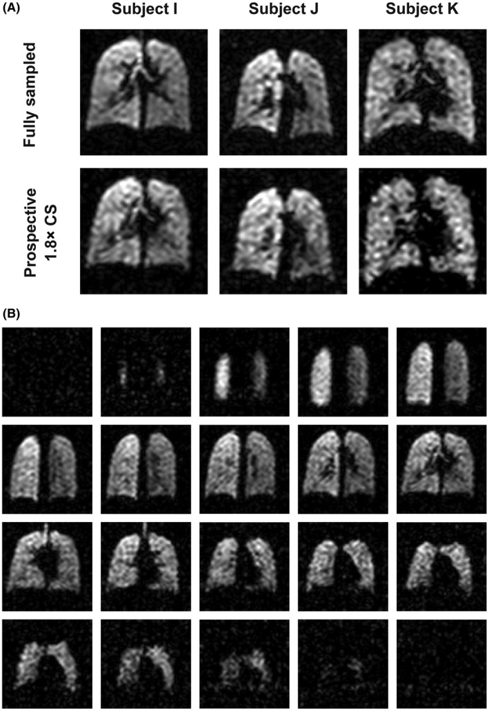
Figure 6.

A, Coronal slices from 3D fully sampled and prospectively 1.8× accelerated scans (NSA = 4), acquired from three healthy volunteers in separate breath holds. Difference images are not shown because of lack of registration between images resulting from minor difference in lung inflation levels between breath holds). B, A 3D 19F‐MRI scan acquired with 1.8× prospective acceleration (NSA = 4) in a scan of 10‐s duration, showing whole‐lung coverage at 1‐cm isotropic resolution; NSA, number of signal averages
