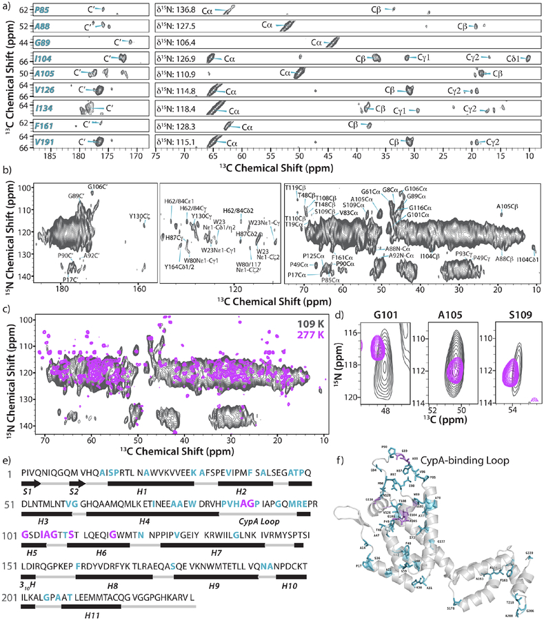
Figure 2.
DNP-enhanced 2D- and 3D NCACX correlation spectra of U-13C, 15N HIV-1 CA tubular assemblies acquired at 14.1 T and 109 K. (a) Selected 2D strips of the 3D NCACX spectrum showing well-resolved resonances and their assignments. (b) 2D NCACX spectrum. The MAS frequency was 12.5 KHz, SPECIFIC-CP time for the NCA transfer was 6.5 ms and the DARR mixing time was 40 ms. (c) Overlay of 2D NCACX spectra acquired under cryogenic temperatures with DNP (14.1 T, gray) and under ambient-temperature conditions without DNP (21.1 T, magenta). (d) An expansion of (c) showing three representative, well-resolved resonances. (e) The primary amino acid sequence of CA with the secondary structure shown. (f) The position of amino acid residues observed and assigned by DNP-enhanced MAS NMR spectroscopy shown in the crystal structure of unassembled full-length CA (PDB: 3NTE). In e)-f), assigned residues are colored cyan and residues for which chemical shift distributions were analyzed are colored magenta.

