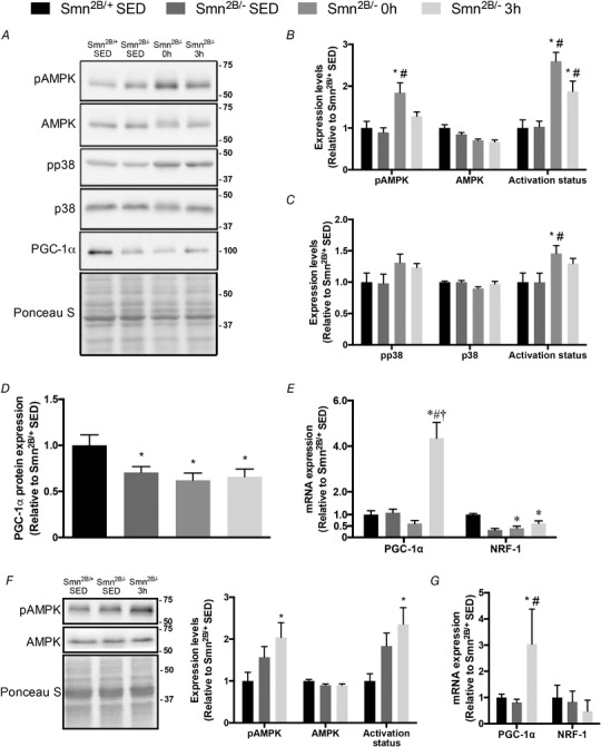
Figure 4. Exercise‐induced signalling in the skeletal muscle of SMA‐like animals.

A, representative western blots of pAMPK, AMPK, pp38, p38 and PGC‐1α in QUAD muscles of Smn2B/+ SED, Smn2B/− SED, Smn2B/− 0 h and Smn2B/− 3 h mice. A Ponceau S stain is also displayed below to demonstrate equal loading across samples. Ladder markers are expressed as kDa. B–D, graphical summaries of pAMPK, AMPK and AMPK activation status (B), pp38, p38 and p38 activation status (C), and total myocellular PGC‐1α levels (D). E, PGC‐1α and nuclear respiratory factor‐1 (NRF‐1) mRNA expression in the TA muscles from all experimental groups. F, representative western blots of pAMPK and AMPK in TA muscles of Smn2B/+ SED, Smn2B/− SED and Smn2B/− 3 h mice. Graphical summaries of pAMPK, AMPK and activation status are shown to the right. G, PGC‐1α and NRF‐1 mRNA expression in the QUAD muscles from Smn2B/+ SED, Smn2B/− SED, and Smn2B/− 3 h animals. Values are expressed relative to Smn2B/+ SED. * P < 0.05 vs. Smn2B/+ SED; # P < 0.05 vs. Smn2B/− SED; † P < 0.05 vs. Smn2B/− 0 h; n = 5–7.
