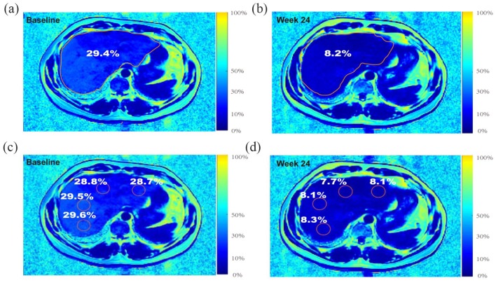
Figure 2.
Example of measuring the percentage change in liver fat fraction with MRI-PDFF. A 45-year-old male with moderate-to-severe steatosis was treated with orlistat. (a) Pretreatment MRI-PDFF demonstrated a total liver fat fraction of 29.4%. (b) The 6-month follow-up MRI-PDFF imaging exhibited a 21.2% decrease in the liver fat content of the total segments of liver (total liver fat fraction of 8.2%). (c) A pretreatment MRI-PDFF map. (d) A 6-month follow-up MRI-PDFF imaging map, placing ROIs centrally in all liver segments for the same patient.
MRI-PDFF, magnetic resonance imaging-derived proton density fat fraction; ROI, region of interest.

