
Figure 1.
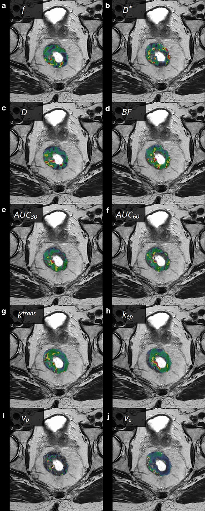
Examples of parametric images from a male patient with a T3 rectal tumor (parametric images of the tumor area shown as color overlays on T2‐weighted images), (a) f, (b) D*, (c) D, (d) BF, (e) AUC30, (f) AUC60, (g) K trans, (h) k ep, (i) v p, and (j) v e.