Figure 8.
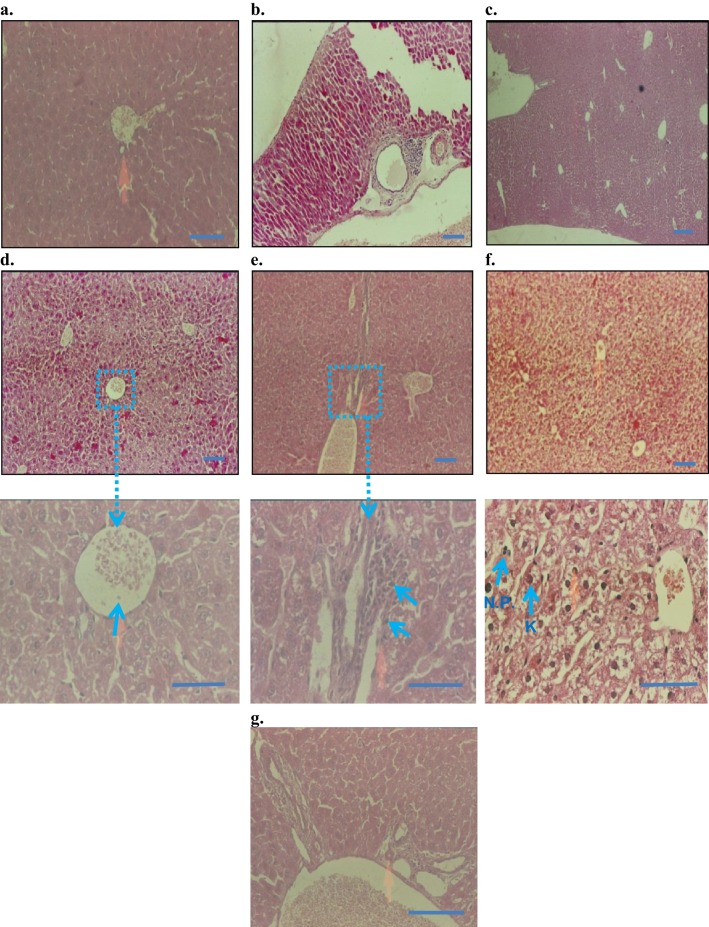
Histopathology of liver (H&E staining, 10× and 50×): (A) control baseline, (B) control injection vehicle, no parenchymal change with very slight periportal inflammation in 10× (Starch-i.p., inj 5 v% for 14 days), (C) no degenerative changes (control paste vehicle), (D) i.p. at 10 mg/kg NP, showing normal histology, (E) i.p. 50 mg/kg NP, slight peritoneal inflammation, (F) i.p.100 mg/kg NP, severe inflammation and liver parenchyma cell death, nuclear pyknosis (arrows), G) topical application 100 mg/kg NP, normal histology, and no signs of inflammation.
Note: Small arrows in (B) and (C) indicate increased amounts of Kupffer cells (K) and nuclear pyknosis (NP). Boxes and connected arrows show the magnification of selected area. Bars 50 µm.

