Figure 2.
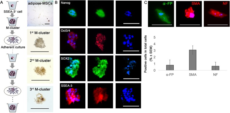
Muse cell characteristics of SSEA-3+ cells sorted from mouse adipose-MSCs. (A) Self-renewal capacity of mouse adipose-Muse cells, as determined by staining for ALP activity in first-, second-, and third-generation M-clusters. Adipose-MSCs were negative for ALP activity. (B) Immunocytochemistry of clusters formed from Muse cells in single-cell suspension cultures, showing that the clusters were positive for Nanog, Oct3/4, Sox2, and SSEA-3. Nuclei were visualized by staining with Hoechst dye. (C) Muse cells in single-cell suspension culture formed clusters in which the cells differentiated spontaneously, expressing endodermal (α-FP), mesodermal (SMA), and ectodermal (NF) markers. Scale bars = 100 µm. α-FP, alpha-fetoprotein; ALP, alkaline phosphatase; M-cluster, Muse cell-derived cell cluster; NF, neurofilament; SMA, smooth muscle actin.

