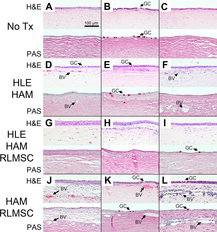
Figure 5.
Basic histology of rabbit corneas at 12 weeks as revealed by staining of sections with hematoxylin and eosin (H&E) and periodic acid–Schiff stain (PAS). Labels “A” through “L” indicate identity of each rabbit as summarized in Table 1. Treatment groups consisted of controls (No Tx), human limbal epithelial cells grown on human amniotic membrane (HLE-HAM), HLE and rabbit mesenchymal stromal cells grown on HAM (HLE-HAM-RLMSC), or HAM with RLMSC alone (HAM-RLMSC). Arrows highlight the location of goblet cells (GC) and blood vessels (BV).

