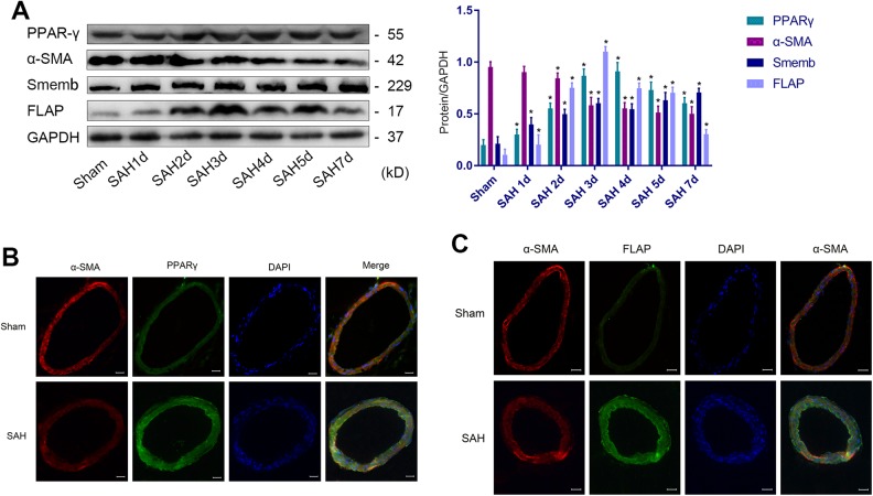
Figure 1.
VSMC phenotypic transformation from 1 to 7 days after SAH. (A) Representative immunoblots and representative quantitative analyses of PPARγ, α-SMA, Smemb and FLAP (*P<0.05 versus Sham group). (B) Representative image and IF of PPARγ (green), α-SMA (red), and DAPI (blue) in cerebral vascular tissues (scale bar, 20 µm). (C) Representative image and immunofluorescence of FLAP (green), α-SMA (red), and DAPI (blue) in cerebral vascular tissues (scale bar, 20 µm; n=12, with 6 used for Western blotting and 6 used for IF).
α-SMA: α-smooth muscle actin; DAPI: 4′,6-diamidino-2-phenylindole; FLAP: 5-lipoxygenase-activating protein; IF, immunofluorescence; PPARγ: peroxisome proliferator-activated receptor gamma; SAH: subarachnoid hemorrhage; Smemb: embryonic smooth muscle myosin heavy chain; VSMC: vascular smooth muscle cell.

