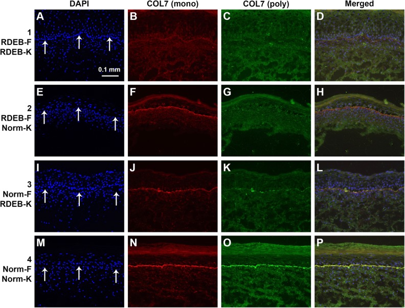
Figure 2.
Localization of collagen VII (COL7) in sections of engineered skin substitutes (ESS) in vitro. Shown are sections of ESS prepared with RDEB fibroblasts and RDEB keratinocytes (group 1; A-D), RDEB fibroblasts and normal keratinocytes (group 2; E–H), normal fibroblasts and RDEB keratinocytes (group 3; I–L), and normal fibroblasts and normal keratinocytes (group 4; M–P). Note that each row depicts a single section photographed using different fluorescent illumination. Immunohistochemistry was performed to localize COL7 using two different antibodies: a monoclonal antibody (B, F, J, N; red), specific for human COL7, and a polyclonal antibody (C, G, K, O; green) that cross-reacts with mouse COL7. Nuclei were counterstained using 4′,6-diamidino-2-phenylindole (DAPI; A, E, I, M; blue). Arrows in A, E, I, and M indicate location of dermal–epidermal junction. Scale bar in A (100 µm) is same for all panels.

