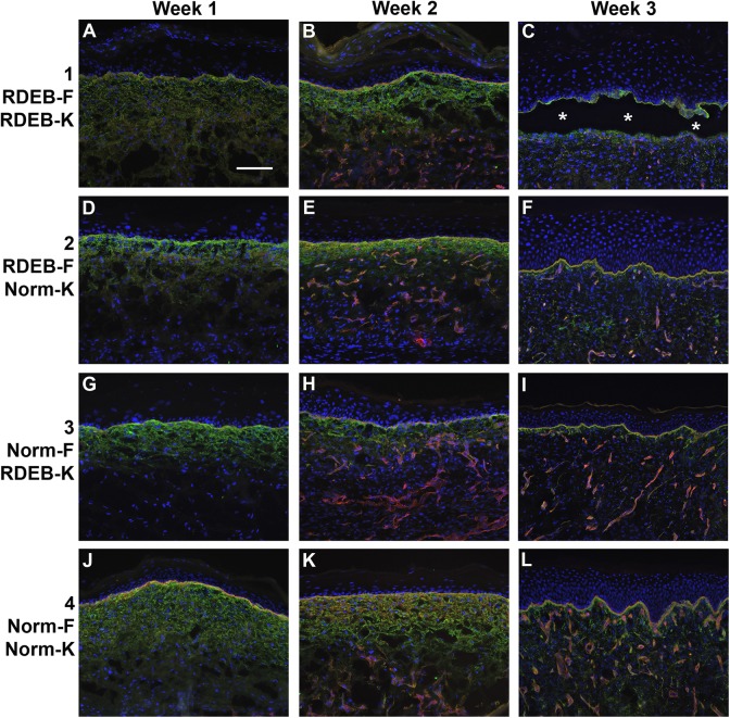
Figure 8.
Deposition of basement membrane in ESS in vivo. Immunohistochemistry was performed to localize collagen IV (green) and laminin (red); nuclei were counterstained using DAPI (blue). Shown are sections of ESS from week 1 (left; A, D, G, J), week 2 (center; B, E, H, K), and week 3 (right; C, F, I, L) in vivo. ESS were prepared with RDEB fibroblasts and keratinocytes (group 1; A–C), RDEB fibroblasts and normal keratinocytes (group 2; D–F), normal fibroblasts and RDEB keratinocytes (group 3; G–I), and normal fibroblasts and keratinocytes (group 4; J–L), as indicated. Asterisks (C) indicate epidermal blistering in ESS prepared with all RDEB cells. Scale bar in A (50 µm) is same for all panels.

