Figure 9.
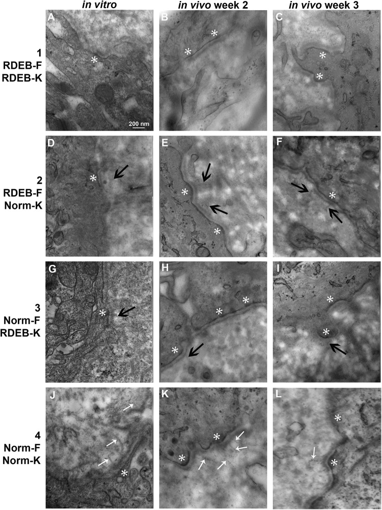
Transmission electron microscopy of dermal–epidermal junction in engineered skin substitutes (ESS). Shown are images of ESS prepared with RDEB fibroblasts and keratinocytes (group 1; A–C), RDEB fibroblasts and normal keratinocytes (group 2; D–F), normal fibroblasts and RDEB keratinocytes (group 3; G–I), and normal fibroblasts and keratinocytes (group 4; J–L), as indicated, collected at the end of the in vitro incubation period (left; A, D, G, J), and at 2 weeks (center; B, E, H, K) and 3 weeks (right; C, F, I, L) after grafting. Anchoring fibrils appeared morphologically normal in samples of ESS prepared with all normal cells (J, K, L; small white arrows); anchoring fibrils in other samples, if present, appeared diffuse and without clear striations (black arrows). Hemidesmosomes were evident in vitro at 2 and 3 weeks in vivo (white asterisks). Original magnification was 80,000×; scale bar in A (200 nm) is the same for all sections.

