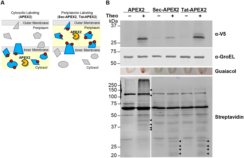
Figure 1. Cytoplasmic and secreted APEX2 have distinct protein biotinylation profiles in Msm.
(A) To localize APEX2 to the periplasmic space, we generated Sec-APEX2 and Tat-APEX2, in which a Sec or Tat secretion pathway signal is fused to APEX2. Red circles denote labeling substrate that reacts covalently with proteins in an APEX2 activity-dependent manner and subsequently serves as an enrichment or detection tag. (B) Msm expressing V5-tagged APEX2 (28 kDa), Sec-APEX2 (31 kDa) or Tat-APEX2 (33 kDa) was grown without or with theophylline and subjected to the labeling protocol with biotin-phenol. Anti-V5 immunoblot analysis of the lysates established expression of APEX2, Sec-APEX2 and Tat-APEX2. Anti-GroEL was used to confirm equal loading. Peroxidase activity was assayed in whole cells using guaiacol. Streptavidin blot analysis was used to detect protein biotinylation. Arrowheads indicate examples of APEX2 expression-dependent bands. Data are representative of >3 biological replicates.

