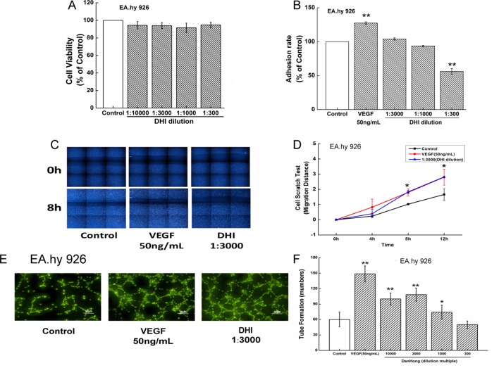
Figure 3.
DHI improved endothelial cells function. (A) EA.hy926 viability was determined by Cell Counting Kit-8 assay under different concentrations of DHI, none dilution caused any significant viability changes. (B) VEGF (50 ng/ml) significantly increased adhesion ability. Lower concentrations (3000, 1000-fold dilution) of DHI did not change the adhesion ability. However, higher concentration (300-fold dilution) of DHI decreased the hy926 adhesion ability compared with the control group. (C) Representative images of the wound healing assay in EA.hy926 cells. (D) Quantitative analysis of the migration distance, VEGF or DHI (3000-fold dilution) promote the cell migration after 8 h. (E) Microscopic images showing tube formation of EA.hy926 cells. (F) After incubating for 12 h with DHI, different dilution folds of DHI promoted angiogenesis in EA.hy926 cells. Data represent the mean ± SD. *P < 0.05, **P < 0.01, compared with the control group.

