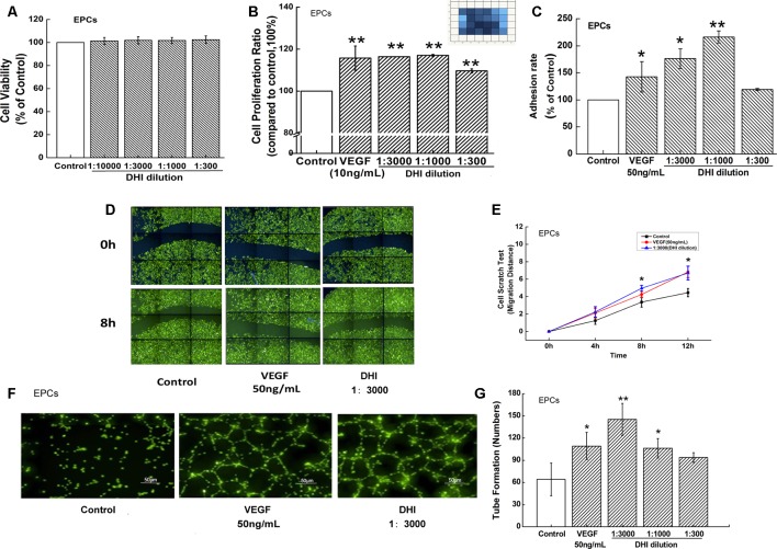
Figure 4.
DHI improved EPCs function. (A) EPCs viability was determined by Cell Counting Kit-8 assay under different concentrations of DHI, none dilution caused any significant viability changes. (B) Different concentrations of DHI significantly increased EPCs proliferation after 48-h cultivation. (C) VEGF (50 ng/ml) and lower concentrations of DHI (3000- and 1000-fold dilution) increased the adhesion ability significantly whereas the highest concentration of DHI (300-fold dilution) had no effect on EPCs adhesion ability. (D) Representative images of the wound healing assay in EPCs. (E) Quantitative analysis of the migration distance, VEGF or DHI (3000-fold dilution) promoted the cell migration after 8 h. (F) Microscopic image showing tube formation of EPCs. (G) After incubating for 12 h with DHI, difference dilution folds of DHI promoted angiogenesis in EPCs. Data represent the mean ± SD. *P < 0.05, **P < 0.01, compared with the control group.

