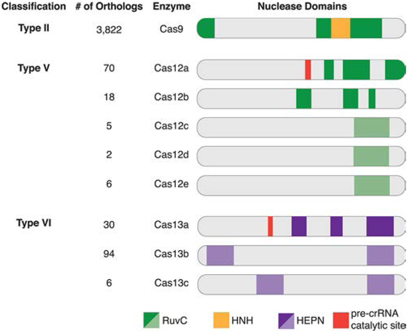
Figure 3.
Schematic representation of the unique Class 2 single effector Cas enzymes and their confirmed or predicted catalytic nuclease domains. Each Cas enzyme is grouped according to its CRISPR-Cas Type classification, and the number of orthologs is listed. The bright colors represent nuclease domains confirmed by crystal structure, and the faded colors represent computationally predicted nuclease domains. Several nuclease domains confirmed by crystal structure are not linear along the amino acid polypeptide chain and are split into sections as shown in the schematic. The split domains include RuvC-like in Cas9, Cas12a, and Cas12b and the first HEPN domain of Cas13a.

