
Figure 1. Widefield calcium imaging during auditory and visual decision making.
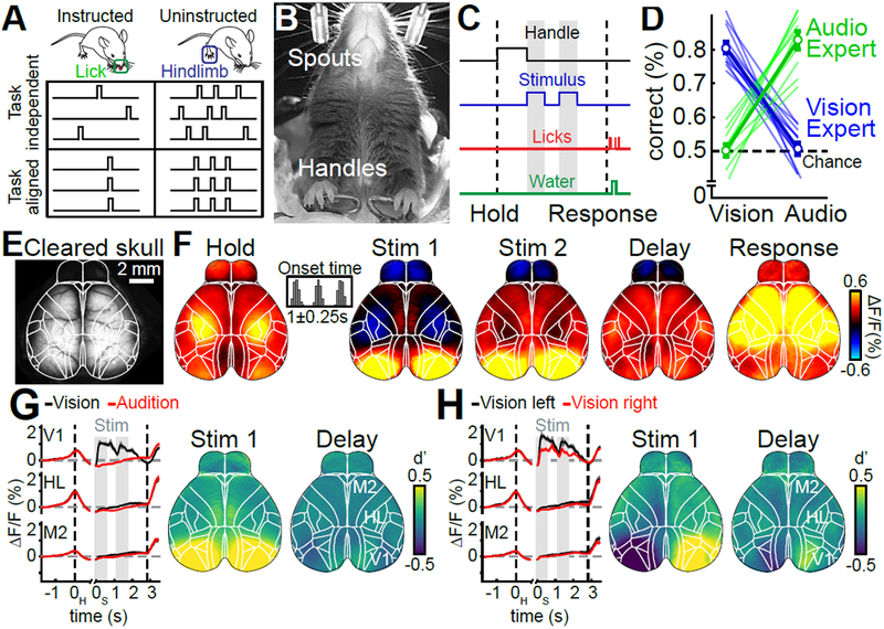
(A) Schematic for the two main questions addressed in this work. Uninstructed movements are exemplified as ‘hindlimb’ but numerous movements are considered throughout this work. (B) Bottom view of a mouse in the behavioral setup. (C) Single-trial timing of behavior. Mice held the handles for 1s (±0.25s) to trigger the stimulus sequence. One second after stimulus end, water spouts moved towards the mouse so they could report a choice. (D) Visual experts (blue) had high performance with visual but chance performance with auditory stimuli. Auditory experts (green) showed the converse. Thin lines: animals, thick lines: mean. Error bars: mean±SEM; n=11 mice. (E) Example image of cortical surface after skull clearing. Overlaid white lines show Allen CCF borders. (F) Cortical activity during different task episodes. Shown are responses when holding the handles (‘Hold’), visual stimulus presentation (‘Stim 1&2'), the subsequent delay (‘Delay’) and the response period (‘Response’). In each trial, stimulus onset was pseudo-randomized within a 0.25-s long time window (inset). (G) Left: Traces show average responses in primary visual cortex (V1), hindlimb somatosensory cortex (HL) and secondary motor cortex (M2) of the right hemisphere during visual (black) or auditory (red) stimulation. Trial averages are aligned to both the time of trial initiation (left dashed line) and stimulus onset (gray bars). Right dashed line indicates response period, shading indicates SEM. Right: d’ between visual and auditory trials during first visual stimulus and the subsequent delay period. (H) Same as (G) but for correct visual trials on the left versus right side. (F-H) (n=22 sessions).