
Figure 2.
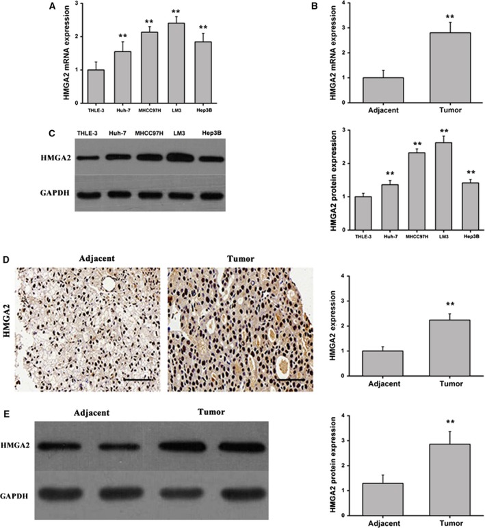
HMGA2 is differentially expressed in HCC cells and clinical cancer specimens. (A) HMGA2 mRNA expression in HCC cell lines (Huh‐7, MHCC97H, LM3 and Hep3B) and normal human hepatocytes (THLE‐3). Data were expressed as the mean ± SD, one‐way ANOVA. (B) HMGA2 expression in HCC tissues and paired adjacent liver tissues. Data were expressed as the mean ± SD, Student's t‐test. (C) HMGA2 protein expression was measured by western blot in HCC cell lines (Huh‐7, MHCC97H, LM3 and Hep3B) and normal human hepatocytes (THLE‐3). Data were expressed as the mean ± SD, one‐way ANOVA. (D) HMGA2 protein expression was measured by immunohistochemical staining and quantitated (scale bars: 50 μm). Data were expressed as the mean ± SD, Student's t‐test. (E) The western blot and quantification of HMGA2 expression in adjacent and tumor tissue. Data were expressed as the mean ± SD, Student's t‐test. n = 6. **P < 0.01.