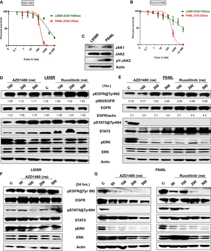
Figure 5.

EGFR variants differ in sensitivity to targeted compounds. (A, B) The sensitivity of EGFR variants (L858R and P848L) to JAK1/2Is (AZD1480 and ruxolitinib) was evaluated using a high‐throughput CellTiter‐Glo. Errors bars represent standard deviation. The specific drug concentrations used were 10 000, 2000, 400, 80, 16, 3.2, 0.64, and 0.128 nm. Briefly, 1000 cells·well−1 were seeded into black clear‐bottom 384‐well plates. Inhibitors were added immediately after seeding. Cells were incubated for 3 days prior to analysis with CellTiter‐Glo luminescent reagent. Plates were read on an M5 Spectramax plate reader; cell viability was normalized to vehicle‐treated wells and fit to a sigmoidal dose–response curve using graphpad prism 6. Experiments were performed in triplicate. (C) Total expressions of JAK1, JAK2, and phospho JAK2 (pY‐JAK2) in Ba/F3‐expressing L858R and P848L were measured by western blotting, and actin serves as loading control. Ba/F3 cells expressing either L858R or P848L EGFR mutation were treated for 1 h. (D, E) and 24 h. (F, G) with the indicated concentrations of JAKi's AZD1480 and ruxolitinib, and autophosphorylations of EGFR at Tyr992, p‐STAT5 at Tyr 694, and p‐ERK were measured by western blotting. Total EGFR, STAT5, ERK, and actin served as loading controls for each experiment. Experiments were done in triplicate, and quantification of phosphorylated EGFR against total EGFR was done using ImageJ software, and using the same software, total EGFR was normalized against actin. Indicated ratios are in arbitrary units. Experiments were performed in triplicate.
