
Figure 8.
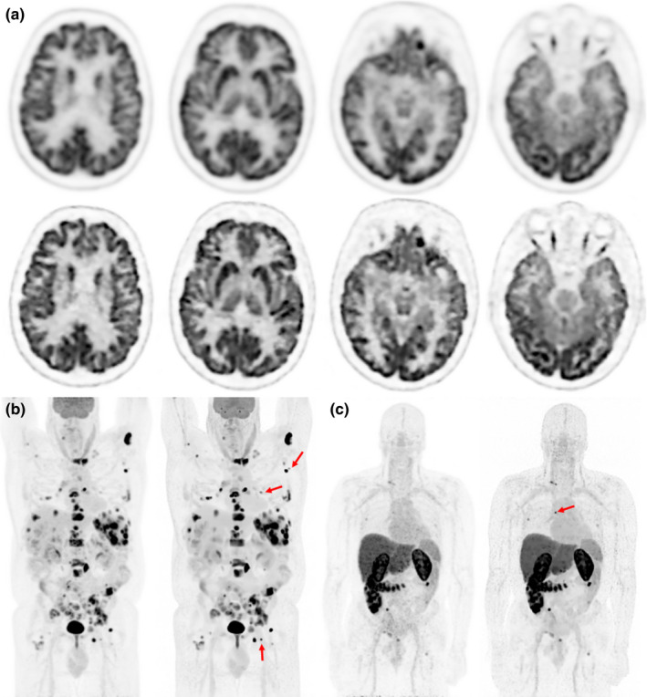
PET Images for three patients are shown. (a) The top two rows show a 18F‐FDG PET brain scan of 52‐yr‐old male with brain metastases. The upper row was reconstructed with our clinical brain reconstruction protocol (postfiltered OSEM) and the lower with ROS‐HOTV‐PAPA. The bottom row show maximum intensity projection images of two different patients, (b) an 18F‐FDG whole body scan of a 59‐yr‐old male with non‐Hodgkin's lymphoma, and C) an Zr‐antibody whole body scan of a 62‐yr‐old male with metastatic prostate cancer, both patients were reconstructed with clinical whole body (postfiltered OSEM) and ROS‐HOTV‐PAPA reconstructions (left and right, respectively). Note the improved sharpness and detail of the brain, and the improved contrast in the whole body and antibody patients’ images. The red arrows indicate lesions that are especially conspicuous in the ROS‐HOTV‐PAPA images. Intrapatient images were identically windowed and leveled. [Color figure can be viewed at wileyonlinelibrary.com]